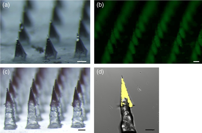
Figure 2.

Micrographs of microparticle‐loaded microneedle patches. (a) Standard patch, (b) fluorescent micrograph of standard patch loaded with fOVA‐loaded microparticles, (c) pedestal patch with sulforhodamine B added to the first PVA/sucrose cast, and (d) confocal image of individual pedestal microneedle containing microparticles loaded with fOVA. Scale = 250 μm
