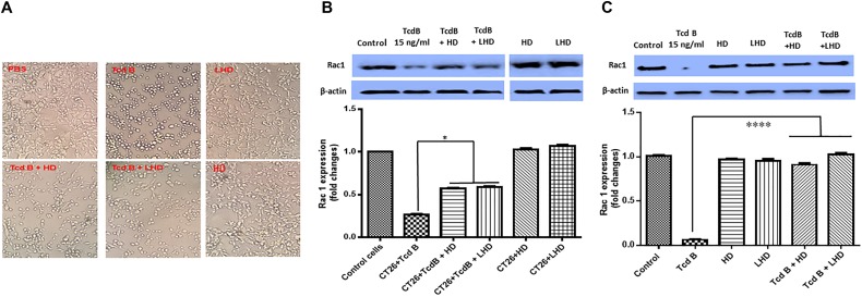
FIGURE 5.
LHD inhibits cytotoxicity of TcdB. (A) LHD inhibits TcdB-induced cell rounding. CT26 cells in 12-well plates were exposed to HD or LHD at 750 ng/ml (50 times of TcdB concentration) or nothing for 1 h, followed by exposure to TcdB at 15 ng/ml for 5 h. (B) Western-blot analysis of non-glucosylated Rac1 in CT26 cells treated with TcdB in the presence or absence of LHD or HD. TcdB glucosylates Rac1 in cells, serving as a readout for toxin cytotoxicity. Up panel shows one blot being cut into two separate blots to remove extra sample lanes. i.e., all bands were from the same blot. Quantitation of Rac1 levels in Western-blot was shown in low panel (∗p < 0.05). (C) CT26 cells were lysed, and the cytosolic fraction was exposed to TcdB (15 ng/ml) with or without HD at 750 ng/ml (50 times of TcdB concentration) or LHD at 1500 ng/ml (same molecular concentration as HD) or nothing for 1 h followed by Western Blot analysis using a monoclonal antibody that only recognizes non-glucosylated Rac1. β-actin was used as an equal loading control. Quantitation of Rac1 levels in Western-blot was shown in low panel (∗∗∗∗p < 0.0001).

