Figure 4.
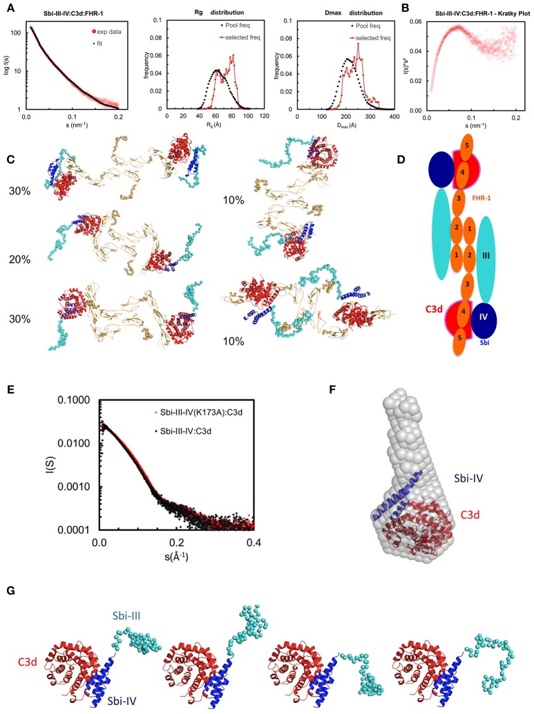
Structural analysis of the Sbi-III-IV:C3d:FHR-1 tripartite complex. SAXS solution structure analysis and EOM modeling of the Sbi-III-IV:C3d:FHR-1 tripartite complex: (A) Left panel, fit of the selected ensemble of conformers to the experimental scattering. Radius of gyration (Rg, middle panel), particle maximum dimension (Dmax, right panel), and distribution histograms of the selected conformers vs. the pool. (B) Kratky plot of the tripartite complex. (C) Examples of rigid body models of the selected conformers corresponding to the histogram peaks. The volume fraction of each species is indicated. The relative positions of C3d, Sbi-III-IV, and FHR-1 in the dimeric tripartite complex are indicated, with C3d in red, Sbi-IV in dark blue, Sbi-III in turquoise and FHR-1 in orange. (D) Schematic representation of the dimeric Sbi-III-IV:C3d:FHR-1 tripartite complex. (E) Comparison of the solutions structure of wild-type Sbi-III-IV:C3d and mutated version Sbi-III-IV(K173A):C3d of the dual complex. Radius of gyration (Rg), particle maximum dimension (Dmax), and distribution histograms of the selected conformers vs. the pool are shown in Figure S4A. (F) Ab initio shape reconstruction shown as gray spheres in comparison to the partial crystal structure Sbi-IV:C3d (2wy8). (G) Examples of rigid body models. Complete set of models as well as flexibility assessment is presented in Figure S5. C3d in shown red, Sbi-IV in dark blue, and Sbi-III in turquoise.

