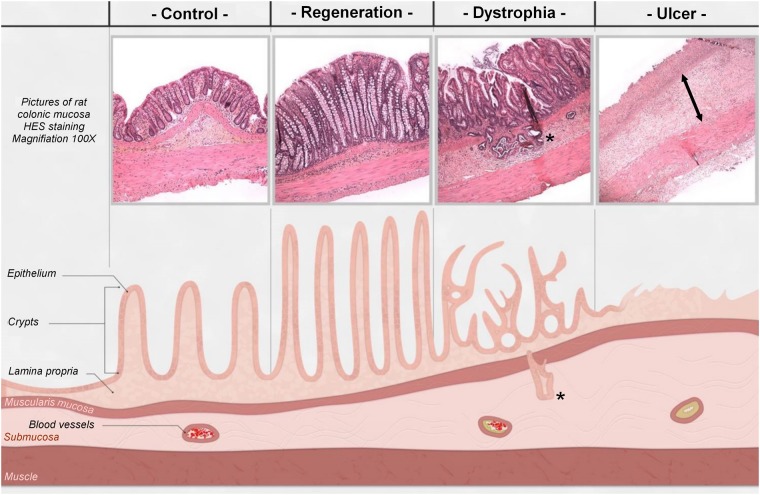
Figure 1.
Representative histological pictures and schematic illustration of the colonic lesion induced after irradiation (4 weeks). In control (non-irradiated) animal, crypts are organized and aligned on the muscularis mucosa. The irradiated area was separated into three zones: (1) The regenerative zone with elongated and organized crypts. (2) The dystrophic zone, characterized by the presence of disorganized crypts with a small edema. Colitis cystica profunda(*) could appear in the most inflamed area. (3) The ulcerated zone where crypts were totally absent and replaced by a dense inflammatory infiltrate with an important edema (<-> arrows) and extracellular matrix remodeling.

