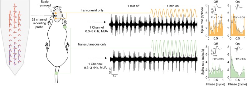
Fig. 2.
Setup to study transcranial- and transcutaneous-only stimulation in rat. A 32-channel silicon probe (far left) was inserted into the motor cortex through a burr hole and used to make extra-cellular recordings of slow “bursting” multiunit activity (MUA, two example recordings are shown in black). A large section of the scalp was retracted allowing transcranial-only sinewave stimulation to be applied through two micro-screws driven halfway through the skull. The orange line shows transcranial-only stimulation. During the experiment, 2-min recordings of MUA were made to compare 1 min with no stimulation (OFF condition), to 1 min with stimulation (ON condition). Transcutaneous-only stimulation (green line) was also a sinewave applied via crocodile-clip electrodes attached to the rectal temperature probe and the skin on one of the four limbs (contralateral fore shown; contra-hind, ipsi-fore and ipsi-hind were also tested). The same 2-min recordings of MUA with OFF and ON conditions were made for transcutaneous-only stimulation. Automatic (Klusta Suite) followed by manual spike sorting was used to isolate single-unit activity. An example of two single-units recorded from the same site are shown in pink and purple on the 32-channel probe. Single-unit spike times were used to calculate cycle histograms (far right panels) to compare the amount of neural entrainment when no stimulation was delivered (OFF, left column) to neural entrainment during stimulation (ON, right column). Neural entrainment in each condition was quantified by calculating the phase locking value (PLV, shown on each cycle histogram). The average single-unit spike waveform for each condition is shown as an inset. In this example, both transcranial-only and transcutaneous-only stimulation cause an increase in neural entrainment from the OFF to the ON conditions

