
Fig. 4.
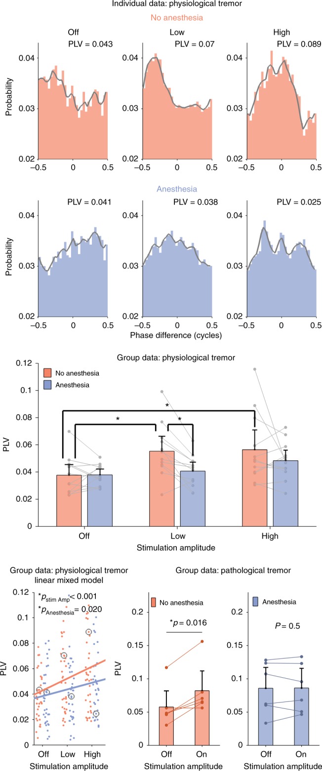
tACS entrainment of physiological and pathological tremor. Effects of focused tACS applied over the motor cortex physiological tremor were tested for three amplitude conditions (OFF, LOW and HIGH) and two topical scalp anesthesia conditions—no anesthesia (blue) and with anesthesia (red). The top two rows show phase-difference histograms and corresponding phase locking values (PLVs) that, in this experiment, quantify the amount of tremor entrainment. The top row shows that increasing tACS amplitude caused an increase in tremor entrainment in one example subject during the no anesthesia condition. The next row shows data from the same subject when the contribution from transcutaneous stimulation of peripheral nerves in the skin was reduced using topical anesthesia. In this condition, increasing stimulation amplitude does not increase tremor entrainment. The gray lines show the smoothed version of the histogram bin values and serve only to highlight the contour. The panel in the third row shows the mean PLV for each condition for all subjects (n = 12). Individual data points (averaged over three repetitions) from the same subject are shown as gray dots and connected with a line. Error bars show the confidence intervals. Significantly different conditions (exploratory post-hoc testing) are indicated with a star. The bottom left panel shows each individual data point (repetitions not averaged), with the gray circles indicating the examples shown in the top two rows. The trend lines were calculated by fitting a linear mixed model to the PLV data and using amplitude and anesthesia as predictors. As expected, increasing stimulation amplitude causes a significant increase in tremor entrainment (p < 0.001). Interestingly, anesthetizing the scalp causes a significant decrease in tremor entrainment (p = 0.020). The center and right panels on the bottom row show data from a similar experiment on 12 essential tremor patients with two amplitude conditions (OFF and ON). In the group that received no topical anesthesia (n = 6, center panel), motor cortex tACS entrained pathological tremor. In the group that did receive topical anesthesia (n = 6, right panel), motor cortex tACS did not entrain pathological tremor (one-sided Wilcoxon signed rank). Individual patient data points (averaged over five repetitions) are shown as dots and lines connect the same subject. Error bars show confidence intervals