
Fig. 6.
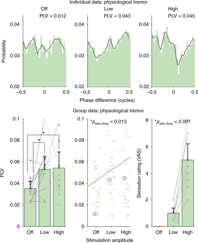
Effect of transcutaneous-only stimulation on physiological tremor. In healthy volunteers, tACS electrodes were placed on the upper arm contralateral to the hand on which tremor was measured, effectively blocking any contribution from a potential transcranial mechanism. The top row shows phase-difference histograms for the three different amplitude conditions (OFF, LOW and HIGH) in one example subject (similar to Fig. 4). Even when the tACS electrodes were not positioned on the head, stimulation still caused physiological tremor entrainment in this subject. The bottom left panel shows the mean PLV for each subject (n = 12) for each amplitude condition. Error bars on all panels show the confidence intervals. Individual data points (averaged over three repetitions) are shown as gray dots. The middle panel shows each individual data point (repetitions not averaged), with the gray circles indicating the examples shown in the top row. The trend line was calculated by fitting a linear mixed model to the PLV data and using stimulation amplitude as a predictor. Even when tACS electrodes are not located on the head, increasing stimulation amplitude still caused a significant increase in tremor entrainment (p = 0.013, linear mixed model). The right panel shows the mean sensation intensity rating for all subjects, using a visual analog scale (VAS) during each amplitude condition. Individual data points are shown as gray dots. The sensation intensity significantly increases with increasing stimulation amplitude (p < 0.001, linear mixed model). This set of data indicate that transcutaneous stimulation of peripheral nerves in the skin can entrain tremor