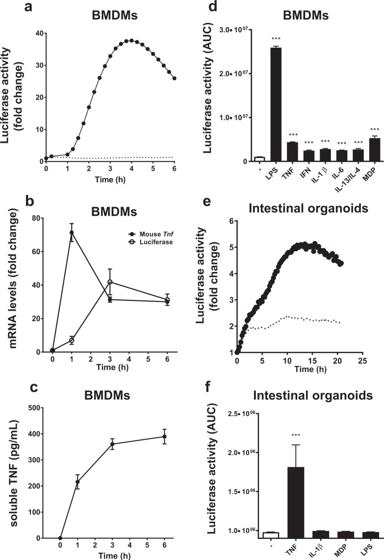
Figure 4.
Responses of in vitro differentiated cell cultures derived from the hTNF.LucBAC mouse strain. Luciferase activity as detected in BMDMs without stimulation (dotted line) or after stimulation (solid line) with 10 ng/mL LPS; data are representative of three independent experiments, with triplicate cultures per experiment (N = 3, n = 3) (a). Tnf and luciferase mRNA abundance as detected in LPS-stimulated BMDMs over 6 h (N = 3 mice) (b). Soluble TNF released to the culture medium in response to LPS treatment of BMDMs over 24 h (N = 6 mice) (c). BMDMs were treated with various ligands and luciferase activity was measured during the cultures (N = 3 mice) (d). Luciferase activity as detected in intestinal crypt stem cell-derived organoids without stimulation (dotted line) or after stimulation (solid line) with 100 ng/mL TNF over 24 h (e). Organoids were treated with various ligands and luciferase activity was measured during the cultures (N = 3 mice) (f); data are representative of three independent experiments, with triplicate cultures per experiment (N = 3, n = 3). Bars represent standard error of the mean. Significant differences from unstimulated controls, **p < 0.01, ***p < 0.001; ANOVA. AUC = area under the curve.

