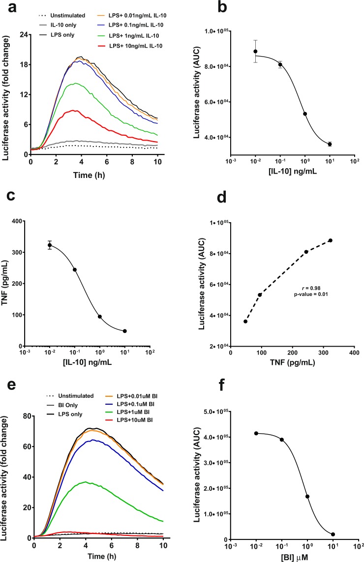
Figure 7.
Effect of anti-inflammatory IL-10 on luciferase activity in hTNF.LucBAC BMDMs. BMDMs were either unstimulated (dotted line) or stimulated with 10 ng/mL LPS, in the absence (black line) or in the presence of increasing levels of recombinant murine IL-10, and luciferase activity was monitored over time; IL-10 concentrations 0.01 (orange), 0.1 (blue), 1 (green) and 10 ng/mL (red) (a). The ED50 of IL-10 as determined by the dose-dependent inhibition of luciferase activity in stimulated hTNF.LucBAC BMDMs was 0.6 ng/mL (b). Soluble TNF detected in the medium 24 h after LPS stimulation in the absence or presence of IL-10 (c). Correlation of IL-10 blockade of LPS-induced luciferase activity represented as area under the curve (AUC) with secreted mouse TNF (d). IKK-2 inhibitor BI605906 (BI) was used as a positive control for inhibition of luciferase activity and soluble TNF release. BMDMs left unstimulated (dotted line) or treated with 10 ng/mL of LPS, in the absence (black line) or presence of BI inhibitor at 0.01 μM (orange), 0.1 μM (blue), 1 μM (green) and 10 μM (red) (e). Luciferase activity (AUC) in the absence or presence of BI in LPS-induced cells (f). Data are representative of three independent experiments, with triplicate cultures per experiment (N = 3, n = 3) and bars represent standard error of the mean.

