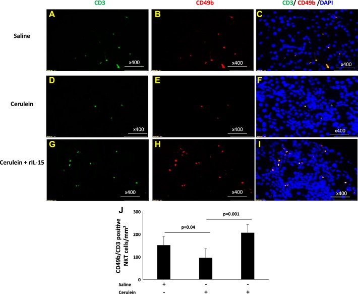
Fig. 5.
Immunofluorescence analysis detected increased natural killer T (NKT) (CD3+CD49b+) cells following interleukin (IL)-15 treatment in cerulein-induced chronic pancreatitis. Double-immunofluorescence analysis was performed to detect CD3+CD49b+ (NKT) cells using anti-CD3-FITC- and anti-CD49b-PE-labeled antibody and mounting with DAPI in the pancreas tissue sections of saline (A–C)-, cerulein (D–F)-, and cerulein with IL-15 (G–I)-treated mice. Morphometric analysis quantitates CD3+CD49b+ (NKT) cells in these tissue sections expressed as NKT cells /mm2 (J). Data are presented as means ± SD; n = 6–8 mice/group. All photomicrographs are shown here with the original magnification ×400. A representative photomicrograph was presented in each group analyzed.

