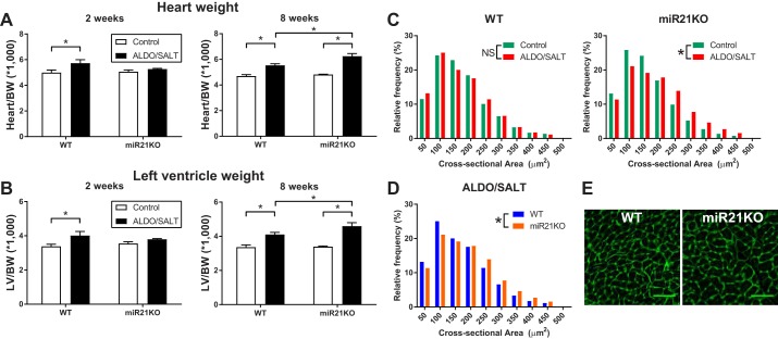
Fig. 2.
miR-21 genetic ablation exacerbates ALDO/SALT-mediated cardiac and cardiomyocyte hypertrophy. miR21KO or WT mice were treated with ALDO/SALT or vehicle for 2 or 8 wk. Heart (A) or left ventricle (B) weights were determined by gravimetry and corrected by body weight. Results are means ± SE (n = 6–8). *P < 0.05. WT or miR21KO mice were treated with ALDO/SALT or vehicle for 8 wk, and cardiomyocyte cross-sectional areas were quantified by WGA staining in FFPE cardiac sections (C and D). Representative images of cardiomyocyte cross-sectional area (E). At least 100 cardiomyocytes in 3 different regions of the right, left, and interventricular septum were quantified, and cardiomyocyte cross-sectional areas were expressed as μm2 (n = 4–5 mice per treatment). Cumulative distributions were analyzed by the non-parametric Kolmogorov-Smirnov test. *P < 0.05. ALDO, aldosterone; BW, body weight; FFPE, formalin-fixed, paraffin-embedded; LV, left ventricle; miR-21, microRNA-21; miR21KO, miR-21 knockout; WGA, wheat germ agglutinin; WT, wild type; NS, not significant.

