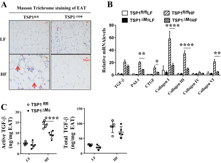
Fig. 5.
High-fat (HF)-fed TSP1∆Mɸ mice had reduced adipose tissue fibrosis compared with HF-fed control mice. A: adipose tissue fibrosis was determined by Masson trichrome staining. The positive staining showed blue color as indicated by the red arrowhead. Representative images are shown. B: expression of fibrosis-related genes in adipose tissue was determined by qPCR and normalized to 18S RNA. C: active and total TGF-β levels in epididymal adipose tissue (EAT) lysates were determined by plasminogen activator inhibitor-1 (PAI-1)/luciferase assay. TSP1, thrombospondin 1. Data are presented as means ± SE (n = 5 mice/group), *P < 0.05, **P < 0.01, and ****P < 0.0001 (2-way ANOVA).

