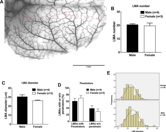
Fig. 6.
Comparison of the structure of leptomeningeal anastomoses (LMAs) from the right rat brain hemisphere from male and female Wistar rats. A: representative image showing the right rat brain hemisphere perfused with carbon black and latex. All identified LMAs were circled in red circles and counted. Scale bar = 5 mm. B: average number of LMAs did not differ between male and female Wistar rats. C: graph showing that average LMA diameter did not vary between male and female Wistar rats. D: graph showing that the percentage of LMAs with or without penetrators did not vary between male and female Wistar rats. E: histogram showing the LMA diameter distribution frequency between male and female rats. No significant difference was observed between male and female Wistar rats.

