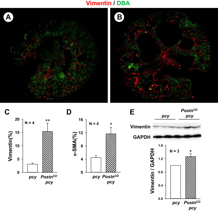
Fig. 3.
Collecting duct (CD)-specific expression of periostin increases the expression of vimentin and α-smooth muscle actin (α-SMA) in pcy kidneys. Representative kidney sections from pcy (A) and PostnCD pcy (B) mice were stained with antibodies to vimentin (red) and DBA (green). There was vimentin staining of mesangial cells in glomeruli in both pcy and PostnCD pcy sections. Cyst-lining cells and interstitial cells of PostnCD pcy kidneys had visually more vimentin staining compared with the kidneys of pcy littermates. Entire kidney sections were imaged using a Nikon Eclipse Ti microscope, and fluorescence for the section was measured using the threshold function in ImageJ software. Bar graphs (means ± SE) represent percentage of positively stained area in tissue section for vimentin (C) and α-SMA (D). E: immunoblots for vimentin and GAPDH levels from 20-wk old pcy and PostnCD pcy kidneys. Bar graph represents the levels of vimentin/GADPH, normalized to pcy (set to 1.0). *P < 0.05, **P < 0.01, compared with pcy mice.

