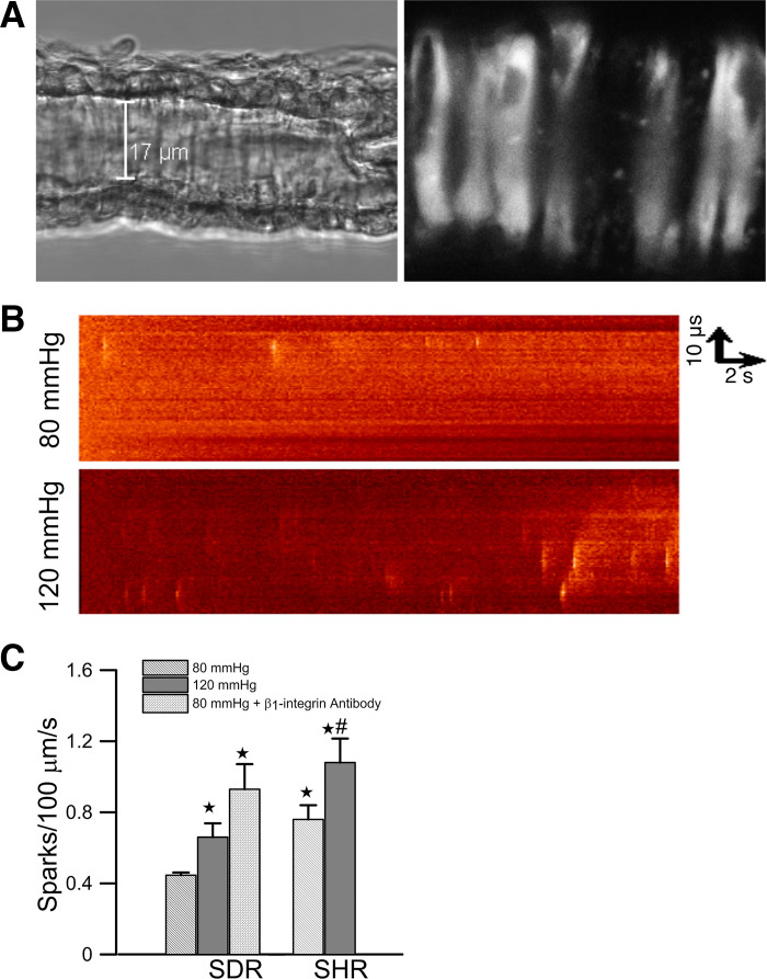
Fig. 1.
A: differential interference contrast image and confocal fluorescence image of an afferent arteriole isolated from a Sprague-Dawley rat (SDR). The arteriole was loaded with fluo-4/AM and perfused at 80 mmHg. Individual vascular smooth muscle cells (VSMCs) are discerned in the fluorescence image. The orientation of the scan line to collect Ca2+ sparks image is shown as a dotted line. Scale bar is 17 µm. B: line scan images of spontaneous Ca2+ spark from VSMCs of afferent arterioles. Images were collected from a single VSMC of an afferent arteriole perfused at 80 mmHg and 120 mmHg, respectively. The images were collected for 16 s from 2 different cells in the same afferent arteriole isolated from a SDR. C: effects of perfusion pressure and β1-integrin activation on the mean Ca2+ spark frequency of afferent arterioles isolated from SDRs (45 cells/10 vessels at 80 mmHg, 50 cells/14 vessels at 120 mmHg, and 13 cells/3 vessels at 80 mmHg in the presence of β1-integrin antibody) and spontaneous hypertensive rats (SHRs) (29 cells/8 vessels at 80 mmHg and 28 cells/6 vessels at 120 mmHg). *Significant difference (P < 0.05) compared with 80 mmHg in arterioles of SDRs. #Significant difference (P < 0.05) compared with 120 mmHg in arterioles of SDRs.

