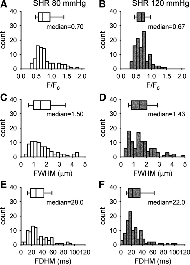
Fig. 3.
The distribution of Ca2+ spark amplitude (A and B), full width half maximum (FWHM) (C and D), and full duration half maximum (FDHM) (E and F) collected from afferent arterioles of a spontaneous hypertensive rat (SHR) perfused at 80 mmHg (29 cells/8 vessels) and 120 mmHg (28 cells/6 vessels). The median of amplitude, FWHM, and FDHM are 0.7, 1.5 µm, and 28 ms at 80 mmHg (n = 84 sparks) and 0.67, 1.43 µm, and 22 ms at 120 mmHg (n = 107 sparks). Box plots show the median and range of the parameters. There is no significant difference in the amplitude, FWHM, and FDHM recorded at 80 mmHg and 120 mmHg.

