Fig. 3.
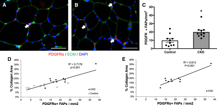
Increased fibrogenic/adipogenic progenitor (FAP) cell abundance within the m. vastus lateralis muscle of CKD patients. A and B: representative immunohistochemical image demonstrating PDGFRα+ muscle FAPs (PDGFRα+ cell surface expression surrounding DAPI+ nucleus; white arrows, red), extracellular matrix (ECM; green) and DAPI (blue) in control (A) and CKD (B) muscle biopsies. Scale bar = 50 μm. C: quantification of FAP content within the muscle represented as mean number of PDGFRα+ FAPs per total muscle area ± SE. D and E: correlation of collagen content within the m. vastus lateralis muscle with FAP content in all participants (D) and CKD patients (E). *P < 0.05.

