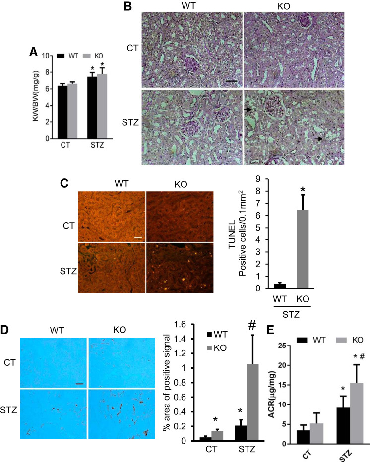
Fig. 1.
Dicer deficiency in proximal tubules aggravates renal tubular injury and interstitial inflammation in diabetic mice. Eight weeks old proximal tubule (PT)-Dicer wild-type (WT) and knockout (KO) mice were treated with streptozotocin (STZ) or without STZ (Control, CT) and examined after 16 wk. A: kidney weight/body weight. Data are expressed as means ± SD. B: representative images of periodic acid-Schiff (PAS) staining showing the vacuolization (arrowheads) in tubules in PT-Dicer KO diabetic. Scale bar: 50 µm. C: representative images of TUNEL assay and TUNEL-positive signal counting showing more apoptotic tubular cells in renal cortex and outer medulla in PT-Dicer KO diabetic compared with PT-Dicer WT diabetic mice. D: representative images of immunohistochemistry staining of macrophage showing more obvious macrophage infiltration in kidney cortex from PT-Dicer KO diabetic mice. E: urine albumin-to-creatinine ratio (ACR). n ≥ 7.*P < 0.05, vs. WT-Control (WT-CT); #P < 0.05, vs. STZ-treated PT-Dicer WT mice.

