Fig. 3.
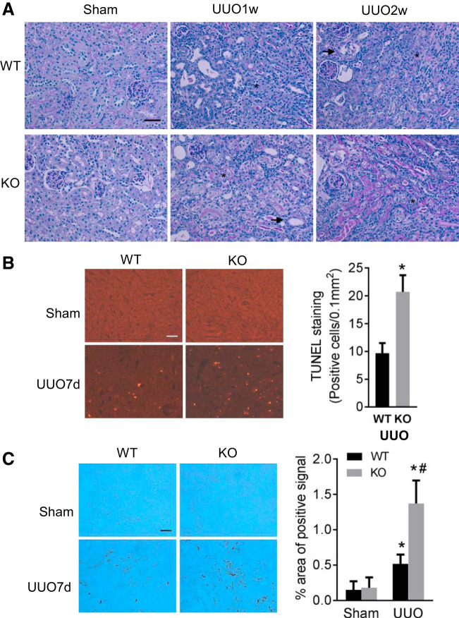
Dicer deficiency in proximal tubules aggravates renal tubular injury and interstitial inflammation in unilateral ureteral obstruction (UUO). Proximal tubule (PT)-Dicer wild-type (WT) and PT-Dicer knockout (KO) mice were subjected to either sham operation (Sham) or UUO surgery. Injured mice were euthanized 1 wk (UUO1w) or 2 wk (UUO2w) after UUO, and left kidneys were collected for histological and immunoblotting analyses. A: representative images of Periodic acid-Schiff (PAS) staining showing more severe renal injury in PT-Dicer KO mice after UUO. Scale bar: 50 µm. Arrowheads and asterisks stand for atrophic and atresic tubules, respectively. B: representative images of terminal deoxynucleotidyl transferase-mediated dUTP nick end labeling (TUNEL) staining and quantification of TUNEL-positive signals showing more apoptotic tubular cells in renal cortex and outer medulla in PT-Dicer KO mice compared with PT-Dicer WT after UUO injury. Scale bar: 50 µm. *Statistically significant difference compared with WT group. C: representative images of immunohistochemistry staining of macrophage showing more macrophage infiltration in kidney cortex in PT-Dicer KO mice compared with WT mice after UUO injury. Scale bar: 50 µm. n ≥ 10. *P < 0.05, vs. WT-sham; #P < 0.05, vs. PT-Dicer WT UUO.

