Fig. 4.
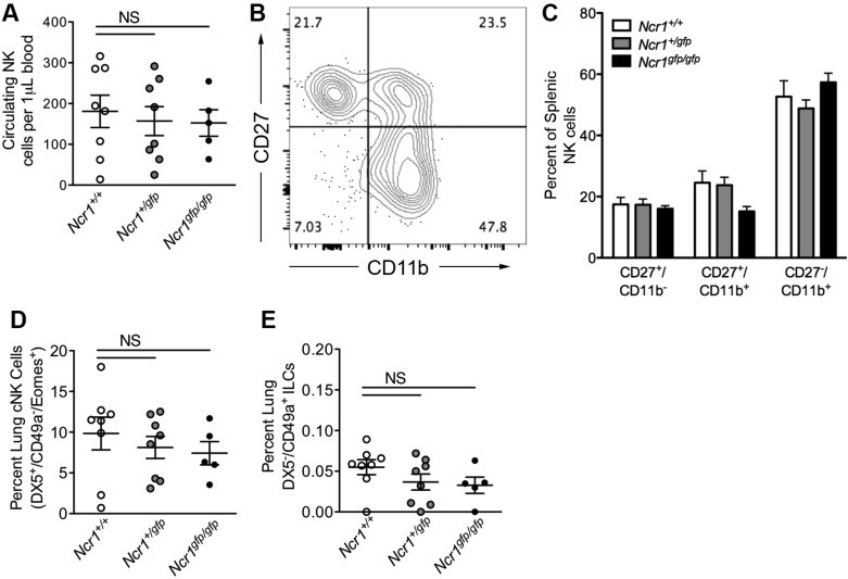
Assessment of natural killer (NK) and innate lymphoid cell (ILC) subsets in the blood and lungs of Ncr1-Gfp mice. A: absolute quantification of circulating CD3−NK1.1+ NK cells in Ncr1+/+, Ncr1+/gfp, and Ncr1gfp/gfp mice (n = 5–8). B: representative flow cytometry plot showing the stages of splenic NK cell maturity, as determined by surface expression of CD27 and CD11b. C: quantification of NK cell maturation in the spleens of Ncr1+/+, Ncr1+/gfp, and Ncr1gfp/gfp mice (n = 3–7). D: quantification of lung conventional NK cells. E: quantification of lung CD49a+DX5− ILC1 cells (n = 5–8). All analyses consisted of one-way ANOVA, Dunnett’s posttest vs. Ncr1+/+ controls. Means ± SE. NS, not significant.

