Fig. 7.
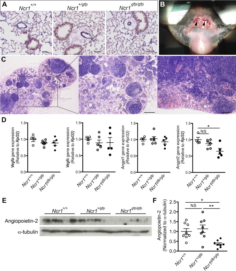
Altered lymphatic function and angiogenic gene expression in Ncr1-Gfp mice. A: representative images of elastic van Gieson-stained fixed-lung sections from Ncr1+/+, Ncr1+/gfp, and Ncr1gfp/gfp mice showing perivascular edema around the pulmonary vasculature of both Ncr1+/gfp and Ncr1gfp/gfp mice. Scale bars = 100 μm. B: image showing enlarged cervical lymph nodes (arrows) in an Ncr1gfp/gfp mouse. C: representative hematoxylin-eosin staining of enlarged lymph nodes from Ncr1gfp/gfp mice. Scale bars = 200 μm. D: assessment of angiogenic gene expression the lungs of Ncr1+/+, Ncr1+/gfp, and Ncr1gfp/gfp mice, normalized to the Rpl32 reference gene (n = 4–6). E: representative immunoblot of angiopoietin-2 protein in the lungs of the mice detailed in A (n = 8). F: quantification of angiopoietin-2 protein in the lungs of the mice detailed in A (n = 8). *P < 0.05 and **P < 0.01, as determined by a 1-way ANOVA with a Tukey’s posttest. Means ± SE.

