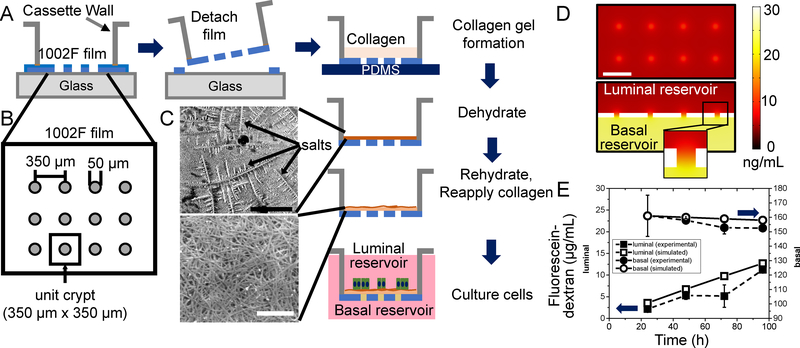
Figure 1.
Fabrication and characterization of the planar-crypt array microdevice. A) Fabrication of the microdevice. Shown is a side view of the 1002F film (blue) patterned with an array of microholes on the surface of a glass slide. After film removal from the glass surface, a layer of collagen (tan) is overlaid onto the 1002F film and dehydrated leaving a compacted collagen layer (brown). After rehydration cells (green) and media (pink) are placed onto the collagen-coated 1002F film. B) Top-view schematic of the film providing the pattern dimensions. A unit crypt is defined as 350 μm x 350 μm square with a microhole in the center. C) Scanning electron micrograph of the top surface of the collagen matrix on the 1002F film before (upper) and after (lower) rehydration. The arrows indicate salt crystals. The black bar (upper panel) represents 200 μm and the white bar (lower panel) 2 μm. D) COMSOL model of the diffusion of a 40-kDa molecule from the basal to the luminal compartment through the microholes. Top panel: Concentration profile of the 40-kDa molecules in the plane just above (10 μm) the luminal surface of the 1002F film. Shown is the concentration profile near 8 microholes in the center of the array at 24 h after addition of the molecule (30 ng/mL) to the basal reservoir. The white scale bar indicates 300 μm. Lower panel: Cross-sectional view through 4 microholes in the 1002F film. Inset shows a cross-section through a single microhole. E) Experimental measurement (solid shapes, dashed lines) and simulation results (open shapes, solid line) of the diffusion of fluorescein-dextran (40 kDa) between the luminal (squares) and basal (circles) compartments over time. At time 0, fluorescein-dextran was added to the basal but not luminal reservoir. The data points represent the average and the standard deviation of the measured data points (n=2) while the lines indicate the simulated concentrations.

