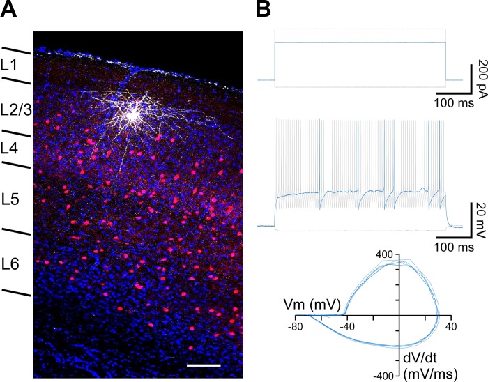
Fig. 1.
Identification of parvalbumin (PV)-expressing interneurons in primary visual cortex using PV-tdTomato mice. A: PV-expressing interneurons were identified by PV-tdTomato (red) expression. Confocal image (×40 magnification) of a recorded PV interneuron stained with biocytin (white), confirming post hoc its location in layers 2/3 of primary visual cortex. DAPI staining (blue) is shown for reference. Laminar distribution is indicated at left. Scale bar, 100 μm. B: the characteristic fast-spiking phenotype of PV interneurons was confirmed for a subset of recordings in current-clamp configuration. Top: applied current stimulation. The PV interneuron was held at Im = 0 pA, and successive pulses of current injection were applied from −50 pA (bottom gray trace) until rheobase (250 pA; blue trace) was reached and up to 350 pA (top gray trace). Middle: membrane voltage responses to the corresponding current injections in top panel (rheobase in blue). Bottom: phase plots of the action potentials evoked at rheobase. The action potentials displayed the characteristic fast kinetics and strong afterhyperpolarization of PV interneurons (Helm et al. 2013; Tricoire et al. 2011). Vm, membrane potential; dV/dt, change in voltage over time.

