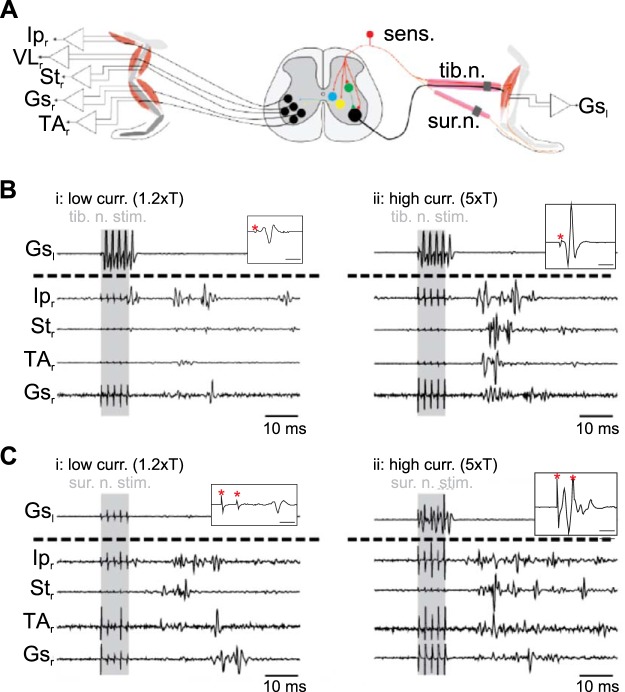
Fig. 1.
Schematic of experimental design used to investigate crossed reflex in vivo. A: experimental design used to investigate crossed reflex in vivo. B and C: examples of electromyographic recording at low (low curr. 1.2×T; i)- and high (high curr. 5×T; ii)-current stimulation from one mouse after stimulation of tibial nerve (tib. n. stim.; B) or sural nerve (sur. n. stim.; C). Shaded areas indicate stimulation. Insets in B and C show examples of the left gastrocnemius (Gsl) response to stimulation of the left tibial nerve with a single impulse (B) or the left sural nerve with double impulses (C). Time bars in insets indicate 2 ms, and red asterisks indicate stimulus artifact. Ipr, right iliopsoas; VLr, right vastus lateralis; Str, right semitendinosus; Gsr, right gastrocnemius; TAr, right tibialis anterior.

