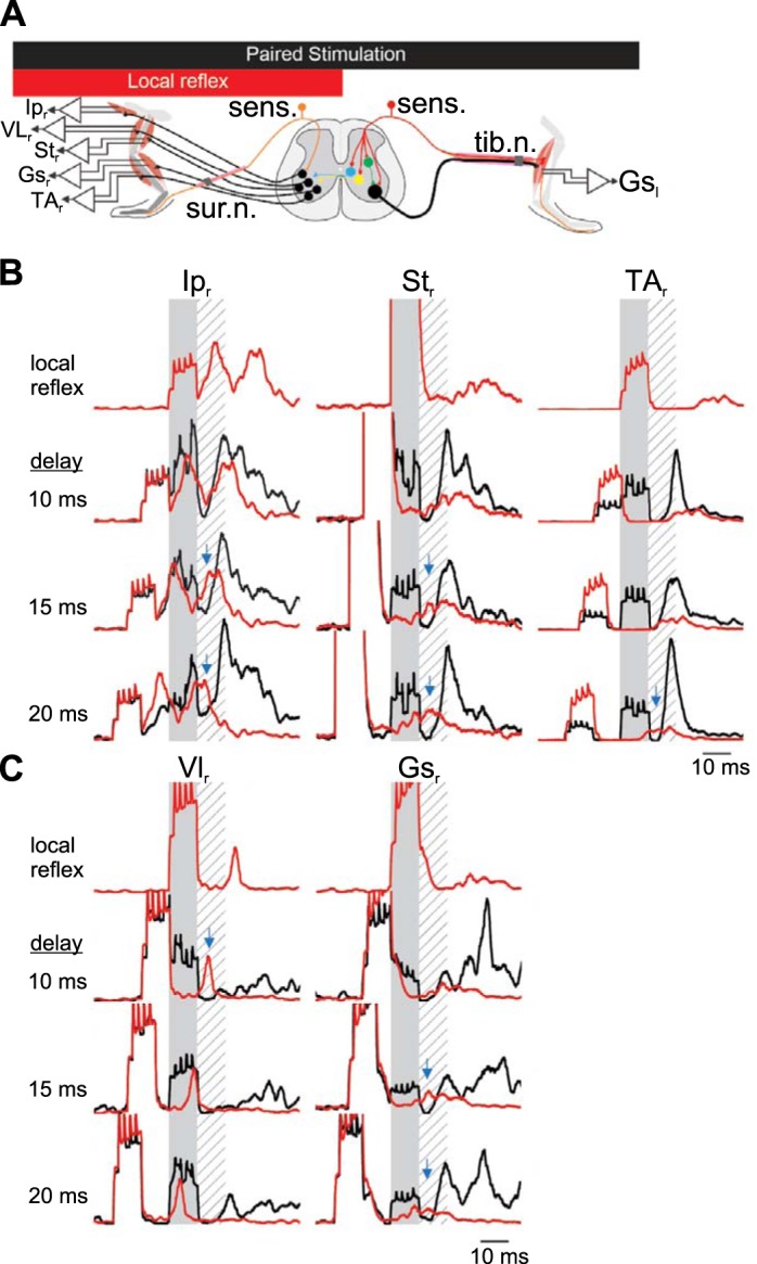
Fig. 5.

Short-latency inhibition in crossed reflex initiated by tibial nerve stimulation. A: schematic representation of the paired stimulation of both legs at different delays to provide evidence of an inhibitory period if there is a suppression of the motor responses from one leg following the stimulation of the other leg. Data are from 40 stimulations from 1 animal, representative of 4 experiments. B and C: examples of electromyographic (EMG) recording in the contralateral flexor muscles (B) or the extensor muscles (C) after stimulation of the contralateral tibial nerve. Shaded areas represent stimulation of the contralateral tibial nerve to evoke a crossed reflex. Hatched areas represent the silent period detected previously. Red traces represent average EMG response to local reflex activation initiated by ipsilateral sural nerve stimulation. Black lines indicate the EMG response when the ipsilateral sural nerve is stimulated with the contralateral tibial nerve with a delay indicated at left of each set of recordings. Blue arrows represent the suppression of the local cutaneous reflex by contralateral sensory stimulation. Gsl, left gastrocnemius; Ipr, right iliopsoas; Str, right semitendinosus; VLr, right vastus lateralis; TAr, right tibialis anterior; Gsr, right gastrocnemius; sens., sensory stimulation; tib. n., tibial nerve; sur. n., sural nerve.
