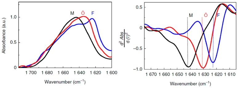
Figure 2.
Left panel: normalized FTIR spectra of Aβ1–40 monomers (M) in 10 mM HEPES/D2O pD11, fibrils (F) after 24 h incubation in 10 mM HEPES pD7.4, and Cu(II)-induced oligomers (Aβ1–40-Cu(II)Õ) after 24 h incubation at 37 °C, pD7.4 [51]. Right panel: second derivatives of the FTIR spectra. Reprinted with permission.

