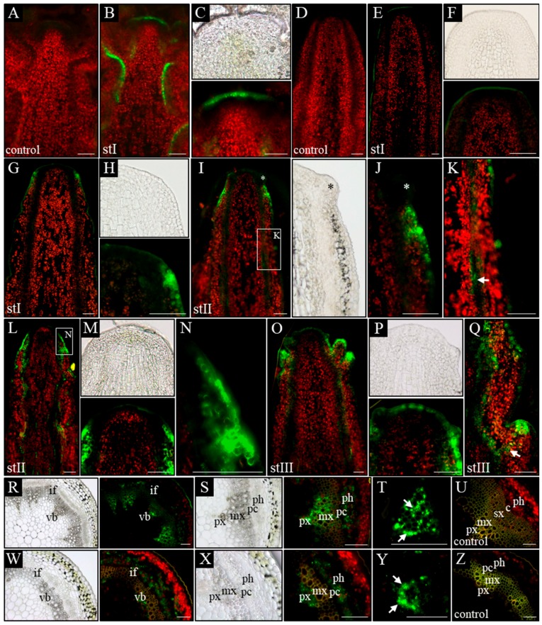
Figure 4.
Spatial localization of the auxin response reporter pDR5:GFP activity. Longitudinal (A–Q) and transversal (R–Z) sections through the apical part of inflorescence stems of WT (A–C,R–U) and pin1 mutant (D–Q,W–Z) plants. On the longitudinal sections, in WT plants, the GFP signal is visible in the vascular strands (B) and the L1 layer of the meristem (C). In the pin1 mutant, in stage I, the activity of the pDR5:GFP transgene is not detected (E,F) or is present in the L1–L2 layers of the peripheral zone of the meristem (G,H); in stage II, GFP signal accumulates in the surface layers at the base of the organ primordia—primordium denoted with asterisk (I,J) and locally in the vascular bundles - denoted with arrow (K) or in the L1–L2 layers of the peripheral zone of the SAM and in all vascular bundles (L–N); in stage III, the GFP signal is visible in the L1 layer of the entire meristem, at the tip of the developing organs and in all vascular strands (O,P) as well as of the cells (denoted with arrow) connecting the tip of an organ primordia with the vasculature (Q). On the transversal sections, the activity of the pDR5:GFP transgene in WT plants is visible in the phloem, xylem and locally in procambial cells (R–U). In terms of the xylem, GFP signal accumulates in all xylem parenchyma cells—denoted with arrows (S,T). In the pin1 mutant GFP signal is visible in xylem and phloem (W–Z). In terms of the xylem, GFP signal is present only in the parenchyma cells on the protoxylem side—denoted with arrows (Y). (A,D,U,Z) Tissue autofluorescence (control); (C,F,H,J,M,P) magnifications of the meristems. St I–III—successive developmental stages; vb—vascular bundle, if—interfascicular fibers, px—protoxylem, mx—metaxylem, sx—secondary xylem, ph—phloem, pc-procambium, c—cambium. Scale bare 50 μm.

