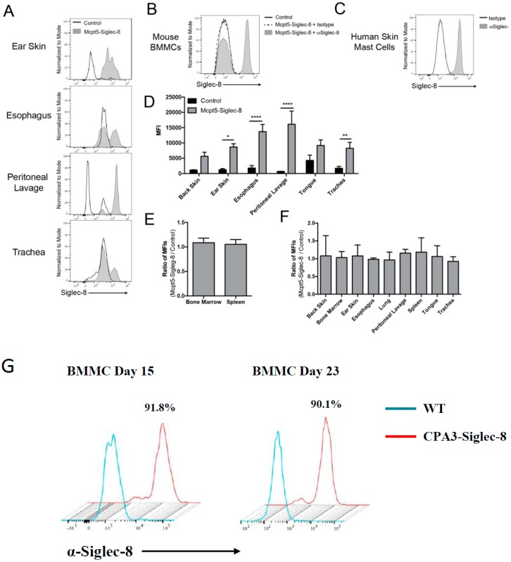
Figure 6.
Mast cells selectively express surface Siglec-8 protein in the Mcpt5-Siglec-8 mouse. (A) Representative histograms of Siglec-8 expression on mast cells (CD45+ CD11b− FcεRI+ c-Kit+ of live cells) in tissues from Mcpt5-Siglec-8 and control mice. Cells from both mice were stained with anti-Siglec-8 mAb (clone 2E2-AF488) and surface expression of Siglec-8 was assessed by flow cytometry; (B) BMMCs from Mcpt5-Siglec-8 and control mice were stained with anti-Siglec-8 mAb or isotype control IgG; (C) Human skin-derived mast cells were assessed for surface Siglec-8 expression; (D) Quantification of MFI of Siglec-8 staining on mast cells (CD45+ CD11b− FcεRI+ c-Kit+ of live cells) isolated from Mcpt5-Siglec-8 (n = 3) and control (n = 4: either WT, n = 1 or Mcpt5-Cre−/− SIG8+/−, n = 3) mice. Ratio of Siglec-8 MFI (Mcpt5-Siglec-8/Control) in non-mast cell populations gated for (E) eosinophils (live CD45+ Siglec-Fmid), and (F) non-immune cells (live CD45−); and (G) surface expression of Siglec-8 on BMMCs from CPA3-Siglec-8 mice. For panels (A–F), shown are data from 3 independent experiments, and the mean±SEM of n = 3–4 are displayed. Shown in panel G are representative flow cytometry plots of two independent experiments. * p < 0.05, ** p < 0.01, **** p < 0.0001 (two-way ANOVA).

