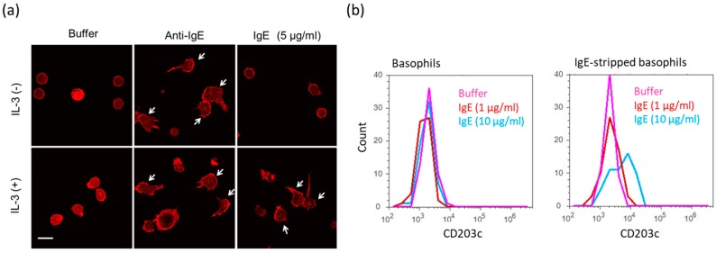
Figure 4.
Various activations of human peripheral basophils in response to high IgE antibody concentrations. (a) Morphological changes of human basophils on the fibronectin-coated glass slide in response to IgE antibodies (5 μg/mL) with or without IL-3. White arrows indicate polarizing cells. White bar shows ca. 10 μm. (b) Expression levels of CD203c on the surface of basophils in response to IgE antibodies (10 μg/mL).

