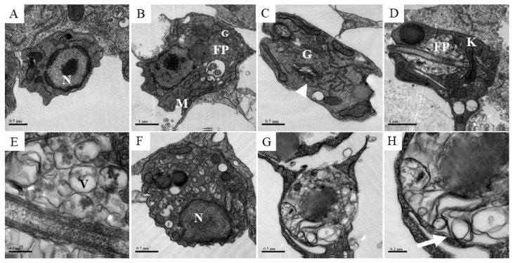
Figure 6.
Ultrastructural analysis of amastigotes after the treatment with compound 2. Peritoneal macrophages from BALB/c mice were infected with stationary phase L. amazonensis promastigotes and treated with compound 2 at 6 µM during 24 and 48 h. (A,B) Control amastigotes showing normal organelles, such as nuclei, mitochondria, flagellar pocket and Golgi. (C,D) Amastigotes treated with compound 2 during 24 h presenting the opening of Golgi (arrowhead) and vesicle accumulation inside the flagellar pocket, respectively. (E) Zoomed image of vesicles inside the flagellar pocket highlighting the double and multiple membrane formation. (F,G) Increased accumulation of cytoplasmic vesicles in amastigotes treated with compound 2 during 48 h. (H) Zoomed image showing a myelin-like figure in the cytoplasm of an amastigote (arrow). N, nuclei; M, mitochondria; FP, flagellar pocket; G, golgi; K, kinetoplast; V, vacuole.

