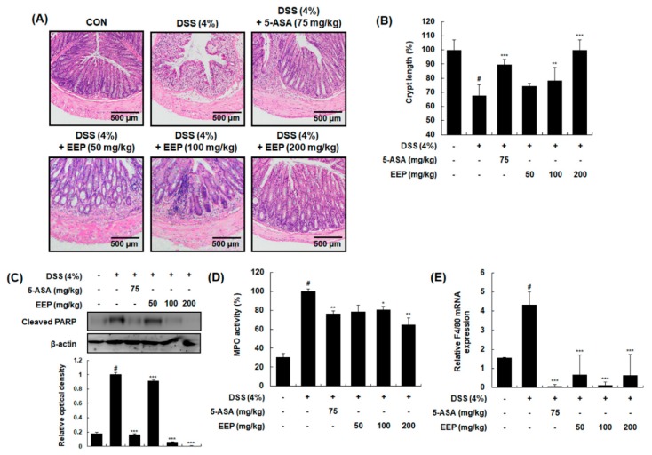
Figure 3.
Effects of EEP on histological changes in DSS-induced colitis mice. (A) Representative sections of colonic tissues from mice administrated 4% DSS in drinking water (ad libitum) for 9 days with or without EEP. 5-ASA (75 mg/kg/day p.o.) was used as a positive control. Histological changes were determined by H&E staining. (B) Changes in crypt length were measured. (C) Whole proteins were prepared from DSS-exposed colonic tissues and subjected to Western blotting to determine expression level of cleaved PARP. β-Actin was used as an internal control. (D) Colon segments from mice were used to determine myeloperoxidase (MPO) levels. (E) Total RNAs were prepared from DSS-exposed colonic tissues and used for qRT-PCR. Using specific primers, mRNA expression levels of F4/80 were determined and normalized against β-actin. Values are presented as means ± SDs (n = 10). # p < 0.05 compared with the vehicle-treated control group, and * p < 0.05, ** p < 0.01, *** p < 0.001 compared with the DSS-induced colitis group.

