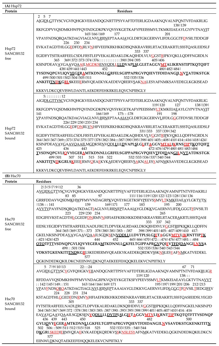
Table 3.
(A and B) Tabulated summary of residues possessing high average BC indices across all ligand-free and ligand-bound trajectories of each protein. Identified residues and respective positions were mapped within the primary sequence. Residues are underlined while positions and numberings are indicated above the residues. Numbering is based on E. coli DnaK template sequence (UniProtKB ID: P0A6Y8). Continuous stretches of residue positions are indicated by colon punctuations. Residues highlighted in red represent residues agreeing with PRS results on E. coli DnaK [18]. Residues existing within the ligand binding site are shown in bold. Corresponding residue numbering from complete sequences of respective proteins are shown in Table S9A,B.

