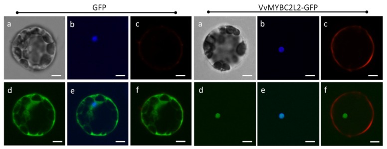
Figure 2.
Subcellular localization of the VvMYBC2L2 protein. Transient expression of the VvMYBC2L2-GFP fusion protein was performed in A. thaliana mesophyll protoplasts. The fluorescent signal was detected by confocal laser-scanning microscopy. Bright field (a), DAPI (b), Dil (Lipophilic membrance dye) (c), GFP (d), GFP + DAPI merge (e), GFP + Dil merge (f). Bars correspond to 10 μm.

