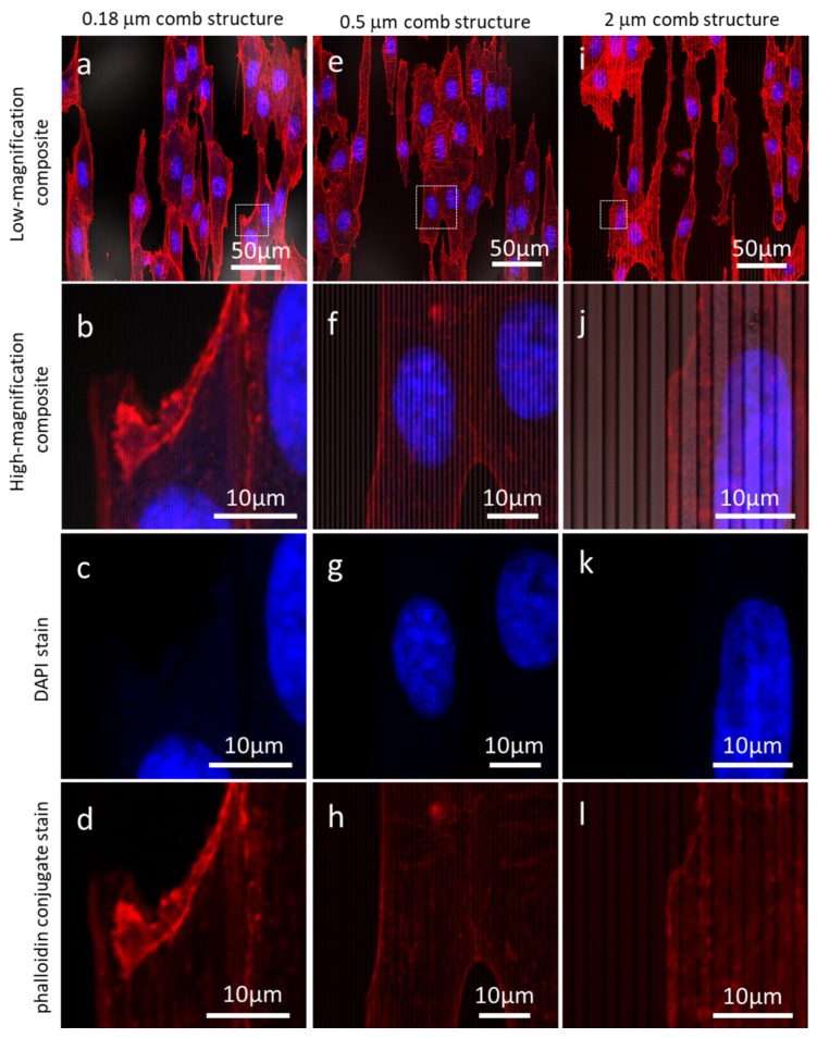
Figure 3.
Low- and high-magnification fluorescence confocal micrographs of cells adhered to 0.18 μm (a–d), 0.5 μm (e–h), and 2 μm comb structures (i–l). The nuclei (blue), actin microfilaments (red) aligned to the pattern axes. Cells were incubated for 24 h with a concentration of ~5 × 10⁵ cells/mL.

