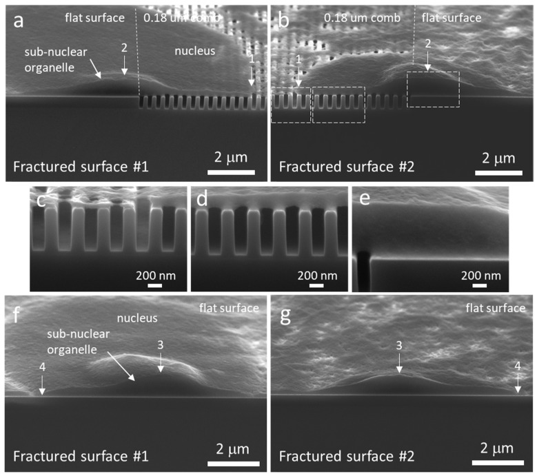
Figure 5.
Tilted SEM micrographs of two cross-sectioned cells. (a–b) Cell adhered on two types of surface (flat and 0.18 μm trench pattern). (c–e) High magnification images of the fractured surface in (b). (f–g) Cell adhered solely on a flat surface. Both sides of the cleaved surfaces are displayed in the figure (left and right columns of images). Landmarks “1–“4” are used to match the topographic features on the two fractured surfaces. In both cells, a micron size sub-nuclear organelle was cross-sectioned.

