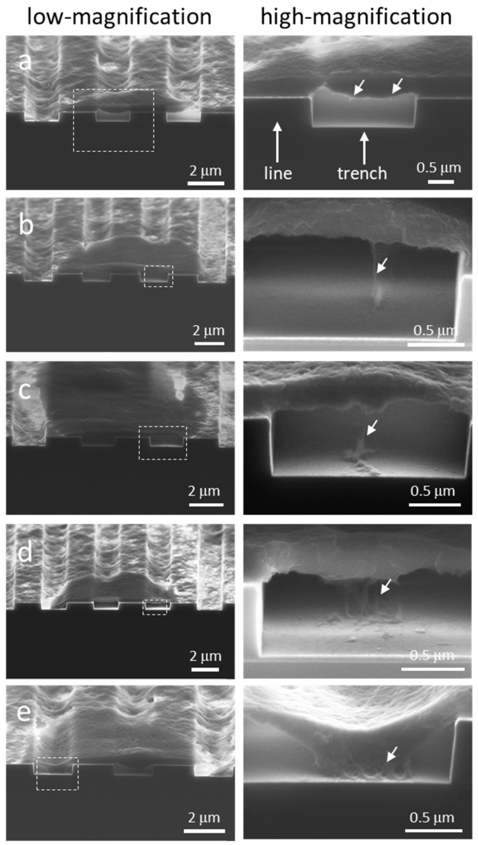
Figure 8.
Micrographs of dissected cells show different amounts of cellular material descending into the 2 μm trenches. The size of filament cluster or fibrous structures that connect the cell underbelly to the substrate increased as more materials sunk into the trench. (a) Short filaments protruding out of the cell underbelly but that did not attach to any surface; (b-c) single-strand filaments from the cell underbelly attached to the bottom of trenches; (d) a cluster of filaments connected the cell underbelly and the bottom of trenches; (e) a large network of fibrous structures is formed between the cell and the trench bottom surfaces.

