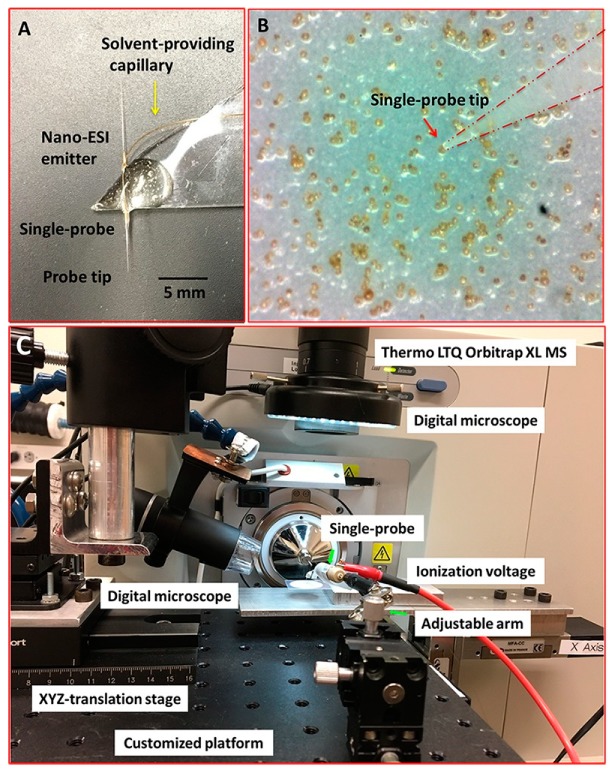
Figure 8.
Experimental setup to measure single Scrippsiella trochoidea cells using the “Single probe” MS techniques. (A) Photograph of the Single-probe device with its different components labeled, including the probe tip and nano-ESI emitter; (B) image from microscope-linked camera used to target single S. trochoidea cell with the Single-probe; (C) setup used to manipulate the Single-probe MS device with components labeled. The Single-probe device was coupled to a Thermo LTQ Obitrap XL MS with an adjustable arm and digital microscope to control transfer of ions entering the MS. Reprinted with permission [12]. Copyright 2018 Sun, Yang and Wawrik.

