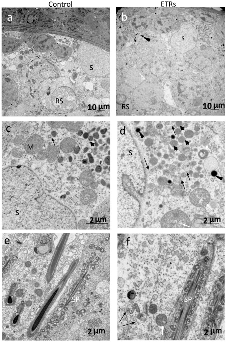
Figure 5.
Ultrastructural features of enhanced autophagy in SCs of ETRs during stages VII–VIII. ((a,c,e): control testes; (b,d,f): ETRs). The long black arrows indicate autophagic vacuoles (AVs) containing membranous structures, while the white arrows mark damaged mitochondria. The short black arrows and arrow heads show lysosomes and lipid droplets (LDs), respectively. RS: round spermatid; S: SC nucleus; SP: sperm; M: mitochondrion.

