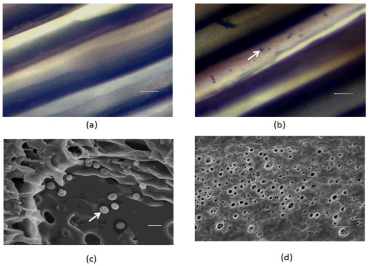
Figure 1.
The light microscopy images of S. epidermidis (a) un-loaded and (b) loaded polysulfone microtube array membranes (PSF MTAMs) and the scanning electron micrographs (SEM) of inner (c) and outer (d) surfaces of S. epidermidis-loaded PSF MTAMs. Arrows indicate the S. epidermidis. Bars (a,b) = 10 µm; and (c,d) = 1 µm.

