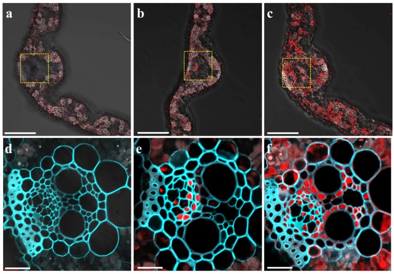
Figure 2.
Tissue-specific and Mg-responsive expression of OsMGT1. Immunostaining with an anti-GFP was performed in the leaf blade of wild-type rice (a,d) and pOsMGT1-GFP transgenic rice under +Mg (b,e) and −Mg conditions (c,f). (d–f) are magnified images of yellow-dotted areas in (a–c) respectively. The red color represents the signal from the GFP antibody and cyan represents the signal from cell wall autofluorescence. Bars = 100 µm (a–c) and 20 µm (d–f).

