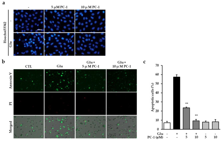
Figure 3.
Procyanidin C1 prevented glutamate-induced apoptosis in HT22 cells. (a) After 12 h exposure to 5 mM glutamate in the presence or absence of 5 or 10 μM PC-1, the nuclei were visualized with Hoechst 33342. Fluorescent images were acquired using a fluorescent microscope. Scale bar, 20 μm. (b) HT22 cells were exposed to 5 mM glutamate in the presence of 5 or 10 μM PC-1 for 10 h and stained with Alexa Fluor 488-conjugated annexin V and PI to evaluate the number of apoptotic and dead cells, respectively. (c) Images were quantitatively analyzed using TaliPCApp software. Bars denote the percentage of annexin V-positive cells (apoptotic cells). Data are presented as the mean value ± S.E.M. ** p < 0.001 versus glutamate-treated HT22 cells.

