Figure 1.
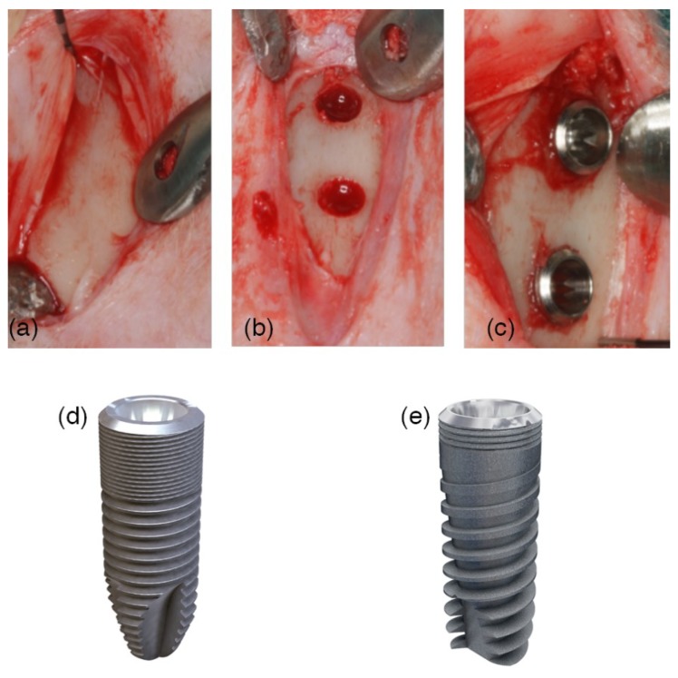
A surgical image. (a) An incision in the skin was performed at the medial section of the tibia. The surgical flaps were raised, showing off the proximal area of the tibia. (b) Two implantation sites were prepared with the same drill protocol, at the metaphysis and the diaphysis areas. (c) Randomly, two implants with different macro-designs were installed and separated by a distance of 8 mm in between. (d) Ticare Inhex®: The implant body had a little conicity and a large area of micro-threads at the coronal portion, and a higher number of triangular threads per unit length and with little thread depth compared to the Quattro® model. Moreover, the implant featured a double self-tapping at the apical portion. (e) Ticare Inhex Quattro®: The implant body had a marked conicity. Fewer micro-threads at the coronal portion and a lower number of macro-threads were present as compared to Ticare Inhex® implants. The threads were squared in the middle part of the implant and became triangular and deeper at the apex, with aggressive self-tapping at the apex.

