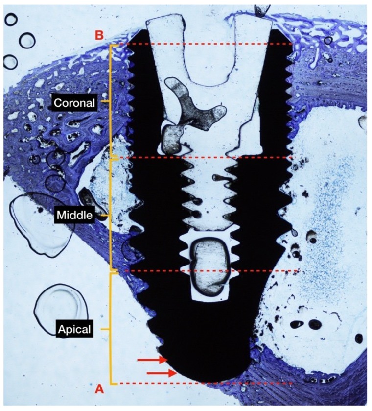
Figure 2.
The ground section of rabbit tibia in diaphysis position at 4 weeks of healing. The implant is divided into 3 equal sections (coronal, middle, apical) for BIC measurement. Two points were traced: (B) The most coronal part of the bone to implant contact, and (A) the base of the implant. The implant surface outside of the cortical bone is not considered in the analysis (red arrows). Original magnification ×2 was used, as was toluidine blue staining.

