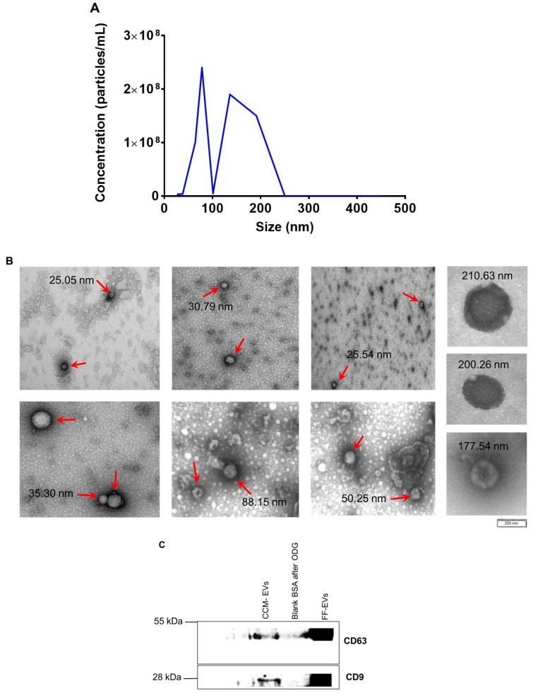
Figure 2.
(A) Nanoparticle tracking analysis, (B) transmission electron microscopy, and (C) western blotting were performed to assess the concentration, size, morphology, and presence of EVs isolated from concentrated conditioned medium (CCM). (A) The concentration of EVs from 1 mL of bovine embryo conditioned medium (obtained from a pool of 500 embryos) was 40.8 × 10⁸ particles/mL and their size ranged between 25 nm and 250 nm. (B) Images demonstrate different EVs indicated with red arrows, size ranging from 25 nm to 250 nm. (C) The presence of EVs in OptiPrep™ density gradient (ODG) ultracentrifugated embryo conditioned medium (fraction 8–9); and blank BSA medium after (fraction 8–9) ODG ultracentrifugation was tested using two EV specific (CD63, CD9). EVs isolated from bovine follicular fluid (FF) were included as positive control respectively.

