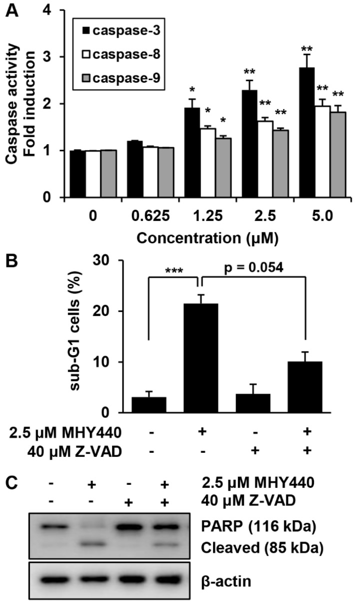
Figure 7.
The effect of caspases on MHY440-induced apoptosis in AGS cells. (A) MHY440-treated cell lysates were assayed for caspase-3, -8, and -9 activities using DEVD-pNA, IETD-pNA and LEHD-pNA substrates, respectively. The emitted fluorescent products were measured. Data are expressed as the means ± SD of triplicate samples. The results represent one of three independent experiments. (B) Cells were pretreated with 40 μM Z-VAD-FMK for 30 min and then treated with 2.5 μM MHY440 for 24 h. Cells were stained with PI and analyzed using flow cytometry. The results are expressed as means ± SD of three individual experiments. Significance was determined using Student’s t-test (* p < 0.05, ** p < 0.01 and *** p < 0.001 compared with vehicle-treated cells). (C) Total cell lysates were prepared and immunoblotted for PARP. β-actin was used as a loading control. Representative results from three independent experiments are shown.

