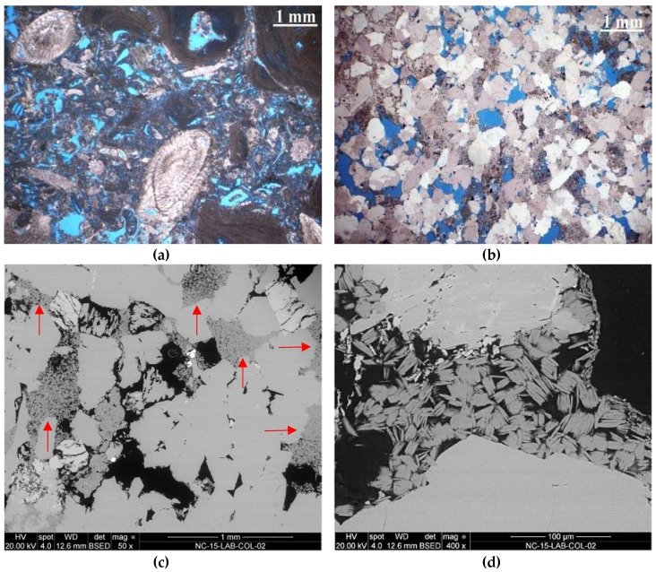
Figure 1.
Microphotos of thin sections recorded with a polarised-light microscope (PLM): (a) St. Margarethen Limestone with different microfossils such as foraminifers, coralline red algae and debris of echinites; (b) Schlaitdorf Sandstone: a coarse-grained quartz arenite with kaolinite and sparitic dolomite; Microphotos of cross sections recorded with a scanning electron microscope (SEM): (c) occurrence of kaolinite (red arrows) up to 15% in Schlaitdorf Sandstone indicating its even distribution throughout the fabric; (d) detail of kaolinite in the intergranular pore space between quartz and feldspar.

