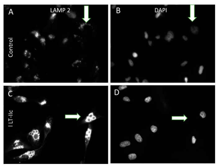
Figure 4.
Enlarged intracellular vacuoles induced by LT-IIc are positive for LAMP-2. (A,B): Untreated MDA-MB-231 cells display punctate cytoplasmic staining for small lysosomes expressing LAMP-2 (A, arrow); DAPI staining for nuclei in same field (B). (C,D): LT-IIc-treated cells at 6 h possess multiple enlarged LAMP-2-stained bodies (C, arrow); DAPI staining of the same field (D). All images were taken under a 40× objective, using identical manual exposure times for control versus treated cells.

