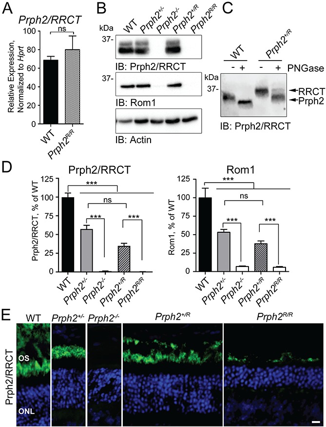
Figure 1.

The RRCT knock-in is expressed in the retina. (A) qRT-PCR was performed on P30 retinal cDNAs using primers that recognize both WT Prph2 and RRCT knock-in transcripts. Values were normalized to Hprt as a housekeeping gene. N = 3 retinas/genotype (mean ± SEM). ns = not significant (P > 0.05) by two-sided Student’s t-test. (B) P30 retinal extracts were separated on 10% reducing SDS-PAGE. Blots were probed with antibodies for Prph2/RRCT (RDS-CT), Rom1 (ROM1-CT) or actin (as a loading control). Shown is a representative blot; blots were repeated with retinal extracts from different animals a minimum of five times. (C) Western blot of untreated and PNGase F treated retinal extracts from WT and Prph2R/R probed for Prph2/RRCT (RDS-CT). (Repeated on extracts from five different animals.) (D) Levels of Prph2/RRCT as percent of WT were measured from the density of the bands in (B) and plotted as means ± SEM from N = 5–10 retinas/genotype. ***P < 0.001, ns: not significant by one-way ANOVA with Tukey’s post-hoc comparison. (E) P30 retinal sections were immunofluorescently labeled for Prph2/RRCT (RDS-CT, green) and nuclei were counterstained with DAPI (blue), images were captured at 40×. OS: outer segment, ONL: outer nuclear layer. Scale bar: 10 μm. Immunofluorescence experiments were repeated at least three times using sections from 3–5 different animals.
