Figure 3.

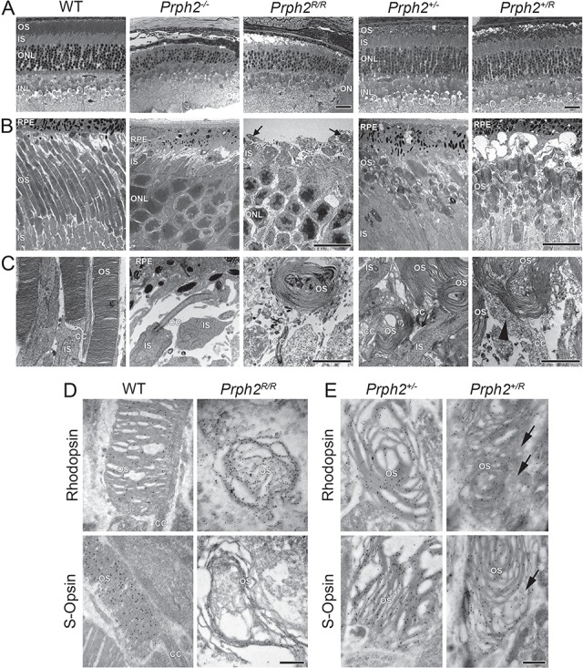
RRCT alone cannot support OS formation while expression of RRCT and WT Prph2 results in accumulation of abnormal vesicular structures. (A) Shown are representative light microscopy images collected at P30. Scale bar: 25 μm. (B and C) Shown are representative P30 EM images collected at 3000× (B) and 15 000× (C). Scale bars: 10 μm (B), 2 μm (C). Arrows indicate tiny nubs of OS in the Prph2R/R, an example of which is shown at higher magnification in (C). Light microscopy and EM were repeated on eyes from at least three different animals. (D and E) Rod OSs were identified by IG labeling with rhodopsin (Fliesler polyclonal) while cone OSs were identified by labeling with S-opsin (Craft polyclonal) antibodies (50 000×). Scale bar: 500 nm. IG labeling was repeated on eyes from at least three different animals. (A–D). RPE: retinal pigment epithelium, OS: outer segment, IS: inner segment, ONL: outer nuclear layer, INL: inner nuclear layer, ON: optic nerve and CC: connecting cilium.
