
Figure 4.
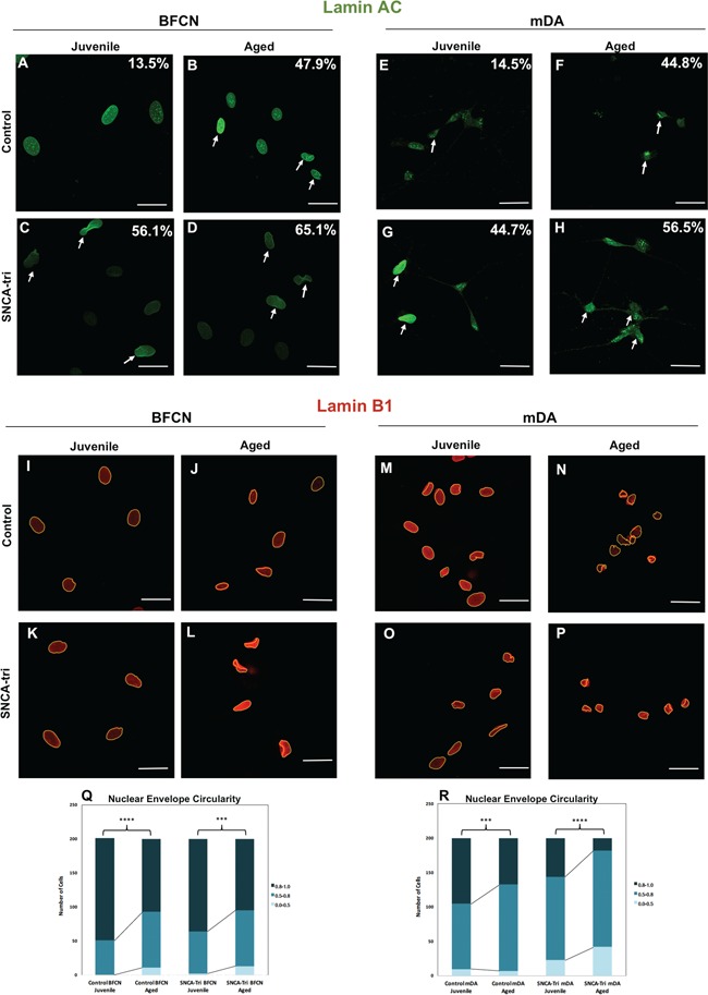
The effects of inducing aging and SNCA multiplication on abnormal nuclear envelope morphology. (A–H) Immunocytochemistry for Lamin A/C in (A–D) BFCN and (E–H) mDA Control and SNCA-Tri lines. Percentages indicate the proportion of cells with folded/blebbed nuclear morphology, white arrows denote abnormal nuclei. Aged hiPSC-derived BFCN and mDA neurons showed higher percentage of abnormal nuclei. SNCA-Tri neurons demonstrated higher percentage of folded/blebbed nuclei compared with the respective Control neurons. (I–P) Immunocytochemistry for Lamin B1 in (I–L) BFCN and (M–P) mDA Control and SNCA-Tri lines. Aged hiPSC-derived BFCN and mDA neurons showed a loss in the nuclear envelope circularity. (Q) Quantification of the nuclear envelope circularity in BFCN demonstrated the loss of nuclear envelope circularity in the Aged versus Juvenile neurons in the Control and the SNCA-Tri lines. (R) Quantification of the nuclear envelope circularity in mDA demonstrated the reduction in the nuclear envelope circularity in the Aged versus Juvenile neurons in the Control and SNCA-Tri lines. A loss in nuclear envelope circularity was observed for the SNCA-Tri lines compared with the respective Control neurons. The data are plotted as frequency distributions of for 200 cells. n = 2 independent differentiation protocols. ***P < 0.001, ****P < 0.0001 according to Kolmogorov–Smirnov test (Q and R). Scale bars: 25 μm. See also Supplementary Material, Tables S17–S19 for all statistical comparisons.